Research Overview
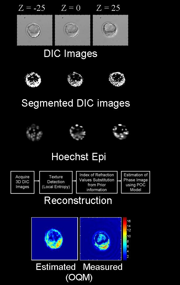
This work introduces a method to extract information about thick transparent samples using multimodality phase microscopy. Phase microscopes are extensively used to image living transparent biological samples because of their ability to obtain high contrast images without the use of enhancing agents. Quantitative phase techniques in particular provide valuable information that can be interpreted easily when the imaged object is optically thin, that is, when the thickness of the object is much less than the depth of field of the imaging system. However, many biological objects of interest have thicknesses comparable to or larger than the depth of field. Our contribution consists of developing a theoretical model based on a product of convolutions (POC) to model phase images of thick transparent objects. The POC model is incorporated in a boundary constrained inversion approach to reconstruct the morphology and indices of refraction of imaged objects.
For more information, contact Heidy Sierra , Charles DiMarzio, Dana Brooks,
Optical Science Laboratory
This research project is part of the work at the Optical Science Laboratory of Chuck DiMarzio in the Department of Electrical and Computer Engineering at Northeastern University. For other projects see Optical Science Lab Research Page.